From the skin to the computer; the data path of electrophysiological data.
1 Introduction
This Whitepaper covers the pathway of electrophysiological data from electrodes on the skin to the computer when measuring with a SAGA amplifier. This pathway is less trivial than might be expected and has a lot of challenges and considerations. This article covers this pathway and provides practical information on the design of a measurement setup.
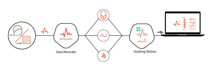
Figure 1: Data path from skin to PC using SAGA.
2 Data path from skin to computer
Generally speaking, acquisition devices capture data. Usually, the data is converted from analogue to digital and transferred to the PC. The conversion of an analogue signal to digital data, AD conversion, consists in essence of taking samples of the signal at (equidistant) points in time. The number of samples taken per unit of time is called the sample rate or sampling frequency, expressed in samples per second or Hertz (Hz). The sample rate is important for various reasons. One of the reasons is to cover the content of the (electro)physiological signal of interest.
The SAGA amplifier consists of a Data Recorder and Docking Station. This design choice has resulted in having a single device that can be applied both in a stationary setup as well as a body-worn setup. Electrodes, placed on the skin, are connected to the Data Recorder. The Data Recorder samples the analogue signal measured at the electrode, resulting in digital electrophysiological data. Hence, the Data Recorder is the acquisition part of the SAGA device. The next step in the signal acquisition pathway is to send the digital signal to the Docking Station. Three interfaces can be chosen to send data from Data Recorder to Docking Station: Electrical, Optical and Wireless. The Docking Station is the device’s means to communicate with a computer. Acquired data can be sent over a USB or network cable to the pc, where the signals measured at the skin can be visualized or processed. Figure 1 shows a graphical overview of SAGA’s data path from skin to computer.
The Docking Station has one more function: Event Triggers can be added to the received data stream. For more information on Event Triggers, please refer to “Knowledge Base article: What are triggers and how to use them?”.
3 Choice of sample rates
Electrophysiological signals (for example EEG and EMG) are complex waveforms. (Electro-)physiological signals have a certain frequency bandwidth and not a single frequency alone (like a sine wave). When electrophysiological signals are measured, you need to take the signal bandwidth into account when choosing the sample rate for acquiring the data. Another important point is whether the Data Recorder can send the acquired data in time to the Docking Station and subsequently to the computer (data transfer bandwidth).
The analysis strategy determines what sample rate should be chosen, but in general one must cover at least the bandwidth of the signal of interest. To cover the complete bandwidth of the electrophysiological signal, ensure that the chosen sample rate is more than two times the highest frequency present in the measured electrophysiological signal.
Another restriction is that the acquisition system also has a maximum bandwidth. SAGA starts measuring at 0 Hz (DC), and it has a maximum analogue bandwidth of 800 Hz at a sample rate of 4 kHz, 700 Hz at a sample rate of 2 kHz, and 350 Hz at a sample rate of 1 kHz.
3.1 Bandwidth of electrophysiological signals
It is important to realize that the limitation of the signal bandwidth, when measuring electrophysiological signals, is determined by the measurement surface and conducting tissue it needs to travel through. It depends on the signal you are measuring and how you are measuring it. This article focuses on surface electrodes, so measuring EMG or EEG from the surface of the skin. This leads to much lower bandwidths of electrophysiological signals compared to invasive measurements. At the skin, we measure the summation of many different action potentials, which are coming from various depths and through fatty or bone tissue. As a rule of thumb, consider the following bandwidth for electrophysiological signals from the body’s surface:
-
- EMG: 10 Hz to 400 Hz
- EEG: DC to 70 Hz (higher frequencies may be relevant for certain applications).
If you want to perform spectral analysis on the EMG signal, the entire bandwidth should be covered to accurately determine the signal’s spectral characteristics. This means that you need to choose a sampling frequency that covers the complete EMG bandwidth. When you are only interested in ‘if’ a muscle is active, you do not need to cover the complete bandwidth necessarily. That behavior can also be captured sufficiently on lower sample rates.
More or less the same is true for EEG signals, although the frequency bandwidth of EEGs is much lower. When you consider this, you might think a sample rate of 500 Hz is already overkill, which in many cases could be true. However, when time resolution is important, it might be just right to sample at higher sample rates.
3.2 Bandwidth of data transfer between Data Recorder and Docking Station
Choosing the highest available sample rate generally results in acquired data with the largest information content. However, choosing a high sample rate results in large amounts of data that need to be transferred from SAGA’s Data Recorder to the PC. When sampling 64 channels with 24 bits per sample at 4000 samples per second, the data rate is 6 Mbit/s. The interface with the lowest data transport bandwidth is the Wi Fi link, which under ideal circumstances runs at 2 Mbit/s. When many channels need to be active simultaneously over the Wi-Fi link, it may be necessary to lower the sample rate to fit the data within the data transport bandwidth.
4 Sample rates per channel type
SAGA supports multiple signal input types: Auxiliary (AUX), Bipolar (BIP) or Unipolar (UNI). These sample rates can be set per signal input type individually.
With SAGA, it is possible to choose a sample rate per channel type or switch off channels that are not used. For example, a low sample rate can be set for AUX channels that are used to measure temperature or acceleration of a body part; properties that usually do not change rapidly. They have a low bandwidth so they can be perfectly sampled at a lower rate. BIP channels on the other hand might be used to measure EMG signals, which can easily reach a frequency content of a few 100 Hz. Hence, this increases the flexibility of the setup for a Wi-Fi measurement; you can design your own channel set configuration to manage data rate and channel setup.
5 Summary
In summary, measuring an analogue signal on the skin and having the signal available on a computer, requires knowledge of the possibilities your data acquisition system offers. Furthermore, knowledge about the frequency content of the physiological signal to be measured is essential in selecting the right measurement configuration. When the maximum data rate is limited (as is the case for the Wi-Fi link) it is still possible to optimize the measurement setup. This is done by selecting different sample rates per signal input type, such that the chosen sample rates cover the entire bandwidth of the different types of signals.